Diabetes and eyes
What is diabetes?
The sugar diabetes is a socially significant disease. It is among the most common chronic conditions, but with the modern methods of diagnostics and treatment it can be monitored and controlled. In a therapeutic aspect, the objective is to maintain the blood sugar within certain limits and to prevent possible complications, thus allowing the diabetic patients to have normal life.
Diabetes is a disease that effects the whole body, and if it is not adequately and properly controlled, it can impair the vision in a way that is difficult to treat. Diabetes-related conditions depend on the duration of the disease, the blood sugar control, and the presence of other concomitant diseases, such as high blood pressure, high cholesterol, overweight, and other conditions.

It occurs in people of different age groups, including people in young active working age. The disease is divided into two main types. Type 1 Diabetes is more typical for young people, while Type 2 affects older people. In diabetes, we can talk about the so-called "rejuvenation" of the disease that is a consequence of the modern lifestyle.

Eye disorders in diabetes
One of the first symptoms is the development at an earlier age of cataract, or the so-called "clouding", which is caused by disrupted metabolism of the natural human lens in diabetes. As a result, the lens clouds and, accordingly, the patients complain of reduced vision. Fortunately, this is a reversible condition with the help of the modern cataract surgery.
However, the main problem is the development of the so-called diabetic retinopathy, which, if poorly controlled and at an advanced stage, can lead to blindness.
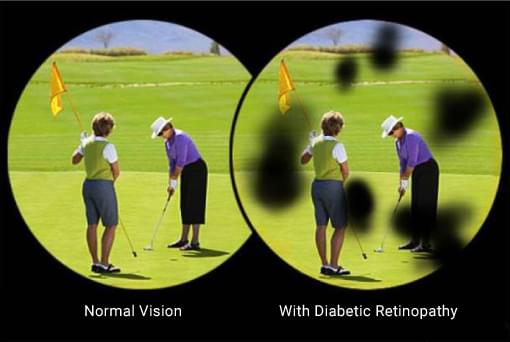
The diabetic retinopathy affects the small blood vessels in the eyes. In this way, the retina, which is the inner sheath of the eye and is responsible for the perception of light, is not properly fed. Damaged vessels can provoke hemorrhages, retention of fluids, which leave the vessels and remain in the retina. The condition is called edema or swelling of the retina. Accordingly, its functions are impaired and the patient's vision decreases.
There are three major types of diabetic retinopathy: non-proliferative retinopathy, maculopathy and proliferative retinopathy.
Typical of the diabetic retinopathy is that at early stage it does not lead to visual impairment, and the patient does not realize that there are changes caused by the disease. It is therefore very important patients diagnosed with diabetes to do periodic preventive examinations. During these exams, initial changes may be detected and, if necessary, they may be treated in order to prevent progression of the condition.

Prevention and tests
Very important for the early diagnosis and the timely treatment of diabetic retinopathy is the patients’ responsibility and their active role in the monitoring of the disease. For those who do not have changes yet, it is enough to have eye examination once a year, including the fundus of the eyes.
When eye changes are detected, fluorescein angiography (color imaging of the eye) is the most appropriate procedure. Fluorescein angiography is performed by intravenous administration of water-soluble contrast. A series of photographs are taken, which show the passage of the contrast agent through the retinal blood vessels, and details of the changes that have occurred as a result of diabetic retinopathy. This study evaluates much more precisely the damage from the diabetic retinopathy, which determines the choice of treatment method.
In case of suspected or already established diabetic macular edema, optical coherence tomography (OCT) is done –computed tomography of the eye. It gives detailed picture of the anatomical changes in the fundus of the eye, and traces with extreme precision even the most discreet tissue changes over time. It is done periodically after treatment.
Treatment
In diabetic retinopathy, "golden standard" for treatment is the so-called laser therapy or ALC. The "color photograph" shows which portions of the retina should be treated with a laser to prevent any progression of the changes. The procedure is done with a device, which looks very much like the device used for examination of the eye fundus. A special magnifying glass is administered to the eye, the procedure lasts 10-15 minutes depending on the area that needs to be treated. There is no pain but minimal discomfort during the treatment. In case of advanced changes of the eye fundus several sessions of treatment may be necessary. The purpose of the laser therapy is not to eliminate the changes caused by the diabetic retinopathy, but to stop their progression. This way the patient's vision is preserved.
Special intraocular medications are applied if there is diabetic edema of the retina, which causes decreased vision. They reduce the edema, help to improve the vision and facilitate the laser therapy.
The advanced changes of the eye in diabetic retinopathy are treated operatively.